


Medical image segmentation using iSeg

Raw MRI data, label-fields, and surface model

Model discretization

Model posing

Illustration of non-manifold triangle meshes (http://pointclouds.org/blog/nvcs/martin/
index.php)

Non-manifold edges occure in situations (b), where more than two regions come together (http://www.cs.utexas.edu/~bajaj/cvc/
software/MMM.shtml)
Model Generation
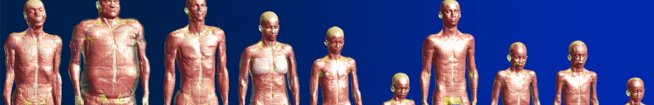
The generation of each anatomical model includes the following 7 steps: 1) recruitment and whole-body scanning of a volunteer, 2) pre-processing of the MRI data, 3) segmentation of the image to generate a label-field, 4) processing of the label-field to remove artifacts, 5) extraction of the tissue surfaces, 6) processing of the surfaces, and 7) quality control. Detailed information is availabe in Gosselin et al. 2014.
Imaging and image pre-processing
- imaging in 1.5 T whole-body scanner
- resolution: up-sampling from 0.5 × 0.5 × 1.0 mm³ in the head and 0.9 × 0.9 × 2.0 mm³ in the torso and limbs (version 1.0) to 0.5 × 0.5 × 0.5 mm³ (version 3.0) by means of the Lanczos interpolation method
Segmentation and label-field processing
- tissue-specific pixel identification with iSEG (view our iSEG training video)
Surface extraction and processing
- extraction and processing of topologically conform triangle surfaces via 1) template-based tetrahedral mesh extraction, 2) surface extraction, 3) feature- and volume-preserving smoothing, and 4) surface simplification
- use of Delauney edge flips to improve mesh quality
Quality control
- use of detailed internal guidelines, including list of necessary structures and tissue definition
- cross-check by 2 or more team members during segmentation and after partial-body and whole-body surface generation
- improvements based on user feedback
Model Processing
Model processing consists of: 1) discretization, i.e., transformation of models into voxels, tetrahedra, etc., for numerical modelling, 2) posing, i.e., posture parameterization, 3) morphing, i.e., changes in a model's morphology, and 4) material assignment. Model posing and morphing are important tools used to extend the population coverage without generating models de novo. Detailed information is availabe in Gosselin et al. 2014.
Discretization
- generation of rectilinear voxel meshes – typically employed for finite-difference techniques – and tetrahedral meshes – commonly employed in finite-element simulations
- generation of conformal sub-cells to reduce the impact of stair-casing possible
- ray-tracing and robust intersection testing used with the rectilinear, non-uniform gridder to create voxel models
- generation of high quality, body-fitted, multidomain tetrahedron meshes with 1) Delauney refinement followed by mesh optimization to remove slivers and improve mesh quality, and 2) a cut-cell octree-based method with a smoothing step
Posing
With the collaboration of SPEAG, users of Sim4Life and Semcad X can now use the Virtual Population as posable models, i.e., model postures can be changed to represent realistic exposure scenarios.
- two approaches are used for posing: 1) a volume-preserving, skeleton- and influence-region-based approach that allows real-time posing and 2) a physical-simulation-based approach that allows the user to first prescribe the position of bones, then performs a tissue mechanics simulation of the passive deformation of the soft tissues, resulting in more-realistic joint-region geometries
- the approaches are implemented with high-performance-computing-enabled solvers, based on the PETSc framework to answer strong computational requirements
Morphing
The morphing functionality allows, e.g., the fat or muscle content to be increased or decreased to change the body-mass index (BMI) of a model while preserving realistic internal organ placement and tissue distribution.
- two approaches are used for morphing: (i) a finite-element, physical simulation-based approach similar to that developed for model posing and (ii) an interactive deformation eld-based warping approach
- approach (ii) has been used, e.g., to enlarge certain organs or to add breathing motion to Virtual Population models
Material assignment
Tissue parameters can be assigned for various physics, e.g., electric and magnetic conductivity, permittivity, and permeability for EM simulations, or thermal conductivity, heat generation rate, heat transfer rate (perfusion), and heat capacity for thermal simulations. Material assignment is based on our database of tissue properties.
Virtual Population V2.0 - supplementary information
The V2.0 surfaces are generated from image segmentations using a technique, which is guaranteed to produce surfaces with compatible interfaces between tissues (triangles are shared between neighboring tissues) and have no self-intersections. These two criteria, as well as good approximation to the segmented labelfields and acceptable triangle element quality have been achieved through dedicated mesh processing algorithms. Please note that compatible surfaces guarantee there are no gaps or overlaps between tissues.
Naturally, since the surfaces are compatible at tissue interfaces, the complete surface (of all tissues) is non-manifold where more than one tissue meets at an edge. Please note, that we cannot guarantee that individual tissue surfaces are manifold.
What does this mean?
- A surface has non-manifold edges if an edge has more than 2 adjacent triangles.
- A surface has non-manifold vertices if the triangles connected to a vertex are not all connected via edges.
