


Figure: Segmentation of brain structures (Manual refinement of automatic segmentation) and vessels.

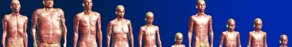
Figure: Frontal and profile view of the Whole-Body model.
ATHENA: 3.5-Year-Old Whole-Body Girl Model
The ATHENA model is a multimodal imaging-based detailed anatomical numerical model of a 3.5-year-old girl. A total of 267 body tissue compartments were segmented, including deep-brain structures, skull, red bone marrow, brain grey matter, brain white matter, skin, subcutaneous fat, cerebrospinal fluid, meninges, muscle, eye, blood vessels, heart, lung, liver, kidney, spinal cord, and cranial nerves. Whole-body MRIs were acquired using MPRAGE, T2 FLAIR, Inversion Recovery, T2 Echo Planar Fast Spin Echo (HASTE), and dual-energy CT scans, which were used to segment the various tissue compartments. An advanced auto-segmentation tool was used on the deep-brain structures.
- ATHENA: 3.5-year-old (Height: 95.4 cm, Weight: 14.7 kg)
- Image modalities used: MRI, CT
- Segmentation resolution: 0.5 x 0.5 x 0.5 mm3
- Number of tissues: 267 tissue compartments in the whole body (including 50 brain tissue compartments)
- Segmented by M.D.s trained for segmentation of pediatric tissues
The ATHENA model can be used for computational modeling studies, such as fluid dynamics, electromagnetics, optics, ultrasound, thermodynamics, and mechanics. Computational modeling with virtual humans helps study the interaction of complex biological problems in silico for source localization, radio-frequency (RF) and specific absorption rate (SAR) exposure, and neurostimulation. The accurate anatomical representation of human numerical models has become integral to many safety studies, such as computed tomography dosimetry and MRI RF exposure.
Important note: The ATHENA model is available in surface (.stl) format. The raw MR and CT image data are not distributed. Please refer to the table below for a list of all the structures included in the ATHENA model.
Version History
| Version | Specifications | Download |
| ATHENA V1.0 |
This model version includes a total of 267 structures. The most detailed image-based anatomical whole-body representation of 3.5-year-old numerical model available for computational modeling studies. |
DOI: 10.13099/ViP-ATHENA-V1.0 License Agreement |
Requests & Inquiries
This model is free of charge for everyone (except for handling fees). To obtain the ATHENA Model, please click on the DOI number above. Please address all inquiries to the Martinos Center or to the Virtual Population Group.
Acknowledgements
The models were developed in cooperation with; the Athinoula A. Martinos Center For Biomedical Imaging, Department of Radiology, Massachusetts General Hospital, Harvard Medical School, Charlestown, MA, USA; the Department of Radiology, Massachusetts General Hospital, Harvard Medical School, Boston, MA, USA; the Fetal-Neonatal Neuroimaging and Developmental Science Center, Boston Children’s Hospital, Harvard Medical School, Boston, MA, USA; and the Center for Devices and Radiological Health, U.S. Food and Drug Administrator, Silver Spring, MD, United States. Financial support was provided by the NIH/NIBB grant R01EB024343.
Reference for citation
A high-resolution pediatric female whole-body numerical model with comparison to a male model. G. Ntolkeras, H. Jeong, L. Zöllei, A. A. Dmytriw, A. Purvaziri, M. H. Lev, P. E. Grand, G. Bonmassar, Phyiscs in Medicine, 2023, doi.org/10.1088/1361-6560/aca950
Disclaimer
The mention of commercial products, their sources, or their use in connection with material reported herein was not to be construed as either an actual or implied endorsement of such products by the Department of Health and Human Services.
