


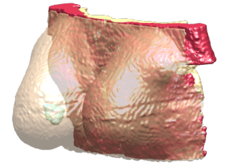
Example model from the Breast Tumor Patient Model Repository (BTPMR).
Breast Tumor Patient Models Repository
Developed at Erasmus University Medical Center, the Breast Tumor Patient Models (BTPM) Repository consists of 22 unique breast models that are segmented into six tissues including a tumor tissue [1]. The main goal of this repository is to create a unique platform to advance the field of intact breast hyperthermia by facilitating an objective comparison of the heating quality for different hyperthermia devices using the same set of patient-derived models. The models in this repository offer a large variability of anatomical and pathological characteristics in the intact breast. Two of the models (Models 4 and 20) are also part of the ESHO benchmarks for computational modeling and optimization in hyperthermia therapy [2].
In terms of anatomical characteristics, the breast models range from small volumes (154 ml) to large volumes (1336 ml) and a mean value (591 ml). Another important anatomical characteristic, the breast density, is well covered, with three out of four breast density groups included in this database. The missing type I (almost entirely fat) breast density category is known to have a significantly lower risk of developing breast cancer, and hence does not appear in this database.
In terms of pathological characteristics, all the repository models hold important variations in terms of tumor size, tumor location, and tumor depth. In terms of tumor size, the tumor volumes vary from very small 1.8 ml to 39 ml. The vast majority of tumors are stage T2 (20–50mm) tumors (18/22), but both T1 (<20 mm; 2/22) and T3 (>50 mm; 2/22) staged tumors are also present in this repository. In terms of tumor location, the current repository covers all the areas of the breast, with the most common tumor site being the upper outer region. In terms of tumor depth, the tumor sites cover all ranges from superficially positioned tumors to tumors very close to the breast base, close to the chest wall. This is evident in the large variation of tumor center to skin distance, varying from 12 to 43 mm, with an average of 26 mm.
All models in the BPTM Repository were created from fully anonymized contrast enhanced MRI data of the breast cancer patients undergoing neoadjuvant chemotherapy. All models were segmented into six tissues: skin, bone, muscle, tumor, fibroglandular, and fat. Skin, thoracic bones, pectoralis major muscle, and the tumor lesion were manually contoured. The remaining tissue volume was segmented into fatty and fibroglandular tissue using an automatic segmentation method based on the voxel intensity described in Zastrow et al. [3] and Omer et al. [4].
Version History
| Version | Specifications | Download |
| BTPMR V1.0 |
Each model contains following tissues. |
DOI: 10.13099/VIP-BTPMR-V1.0 License Agreement |
Requests & Inquiries
This model is free of charge for everyone (except for handling fees). To obtain the BTPM Repository, please click on the DOI number above. Please address all inquiries to the Erasmus Medical Center, Department of Radiotherapy, Hyperthermia Unit or to the Virtual Population Group.
Acknowledgements
This work has received funding from the European Union’s Horizon 2020 research and innovation programme under the Marie Sklodowska-Curie Grant agreement no. 845645.
Reference for citation
Patient-derived breast model repository, a tool for hyperthermia treatment planning and applicator design
Androulakis, I., Sumser, K., Machielse, M.N., Koppert, L., Jager, A., Nout, R., Franckena, M., van Rhoon, G.C. and Curto, S., 2022. International Journal of Hyperthermia, 39(1), pp.1213-1221.
Additional References
[1] Androulakis, I., Sumser, K., Machielse, M.N., Koppert, L., Jager, A., Nout, R., Franckena, M., van Rhoon, G.C. and Curto, S., 2022. Patient-derived breast model repository, a tool for hyperthermia treatment planning and applicator design. International Journal of Hyperthermia, 39(1), pp.1213-1221.
[2] Paulides, M.M., Rodrigues, D.B., Bellizzi, G.G., Sumser, K., Curto, S., Neufeld, E., Montanaro, H., Kok, H.P. and Dobsicek Trefna, H., 2021. ESHO benchmarks for computational modeling and optimization in hyperthermia therapy. International Journal of Hyperthermia, 38(1), pp.1425-1442.
[3] Zastrow, E., Hagness, S.C. and Van Veen, B.D., 2010. 3D computational study of non-invasive patient-specific microwave hyperthermia treatment of breast cancer. Physics in Medicine & Biology, 55(13), p.3611.
[4] Omer, M. and Fear, E.C., 2018. Automated 3D method for the construction of flexible and reconfigurable numerical breast models from MRI scans. Medical & biological engineering & computing, 56(6), pp.1027-1040.
