



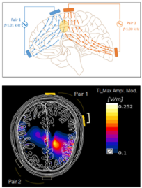
Simulated envelope modulation magnitude map of two interfering E-fields during temporal interference stimulation (Cassarà et al., 2025a, 2025b).
Exposure and EM-Neuro Modeling
Physics-based EM Field Modeling
We quantify electric and magnetic in vivo exposure conditions from desired sources (e.g., transcranial, transcutaneous, and implanted stimulators) and undesired sources (e.g., MRI gradients and other low-frequency fields). Anatomy-accurate Virtual Population (ViP) reference models, subject-specific whole-body anatomies, and image-derived head/spine/peripheral-nerve models allow us to analyze realistic exposure scenarios and their neural activation potential.
Coupled EM-Neural Modeling Across Scales
We combine EM field simulations with detailed neural models (axonal, cellular, and population-level) to bridge the gap from physical fields to excitability, timing bias, and mechanistic hypotheses about synchronization and network modulation (e.g., λ-E approximations; axonal and population-level pipelines).
Focus Areas
- Neural exposure modeling: Integrating detailed neuron models with EM simulations to quantify activity (Botzanowski et al., 2022).
- Mechanistic links across scales: Developing coupled EM-neural pipelines to connect exposure to function (Karimi et al., 2025b).
- Open, interoperable tooling: Workflows within our modeling ecosystems (e.g., Sim4Life, BraiNN, o²S²PARC) support FAIR data practices for collaborative, multi-site studies (Osanlouy et al., 2021).
Selected Achievements
- ViP “neuro-functionalized”: Integration of dynamic neuron models into ViP anatomies to predict nerve stimulation, enable individualized head/spine modeling, and support safety and targeting evaluations.
- Standardization: Active contribution to international committees, helping to define and refine exposure safety standards that protect against undesirable neurostimulation (Neufeld et al., 2016a, 2016b).
References
Botzanowski, B. et al., 2022. Noninvasive stimulation of peripheral nerves using temporally‐interfering electrical fields. Adv. Healthc. Mater. 11, 2200075. https://doi.org/10.1002/adhm.202200075
Cassarà, A.M. et al., 2025a. Recommendations for the Safe Application of Temporal Interference Stimulation in the Human Brain Part I: Principles of Electrical Neuromodulation and Adverse Effects. Bioelectromagnetics 46, e22542. https://doi.org/10.1002/bem.22542
Cassarà, A.M. et al., 2025b. Recommendations for the safe application of temporal interference stimulation in the human brain part II: Biophysics, dosimetry, and safety recommendations. Bioelectromagnetics 46, e22536. https://doi.org/10.1002/bem.22536
Karimi, F. et al., 2025b. Precision non-invasive brain stimulation: an in silico pipeline for personalized control of brain dynamics. J. Neural Eng. 22, 026061. https://doi.org/10.1088/1741-2552/adb88f
Neufeld, E. et al., 2016a. Functionalized anatomical models for EM-neuron interaction modeling. Phys. Med. Biol. 61, 4390–4401. https://doi.org/10.1088/0031-9155/61/12/4390
Neufeld, E. et al., 2016b. Investigation of assumptions underlying current safety guidelines on EM-induced nerve stimulation. Phys. Med. Biol. 61, 4466–4478. https://doi.org/10.1088/0031-9155/61/12/4466
Osanlouy, M. et al., 2021. The SPARC DRC: Building a resource for the autonomic nervous system community. Front. Physiol. 12. https://doi.org/10.3389/fphys.2021.693735


